

Fetal Growth Restriction: when the baby stops growing... even at a normal weight.

Dr. André Miyague palestrante na SOGIPA
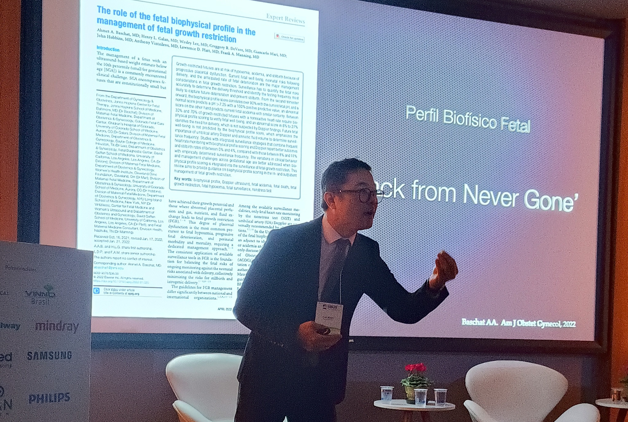
Folks, when we talk about Fetal Growth Restriction, it's very common to only think about that visibly small, thin baby with a lower than expected weight. But the truth is that the case isn't always so obvious. I had the opportunity to speak about this topic at the VI Ultrasound Conference of the SBUS of Paraná – SOPRUS 2025, here in Curitiba. And wow... it was special to be able to share this subject with colleagues, a subject that is increasingly appearing in our daily practice. Today, I want to share some important points from the lecture with you. Because understanding how FGR works — especially late-onset FGR — can completely change the outcome of a pregnancy.

So, what exactly is fetal growth restriction?

This occurs when the baby is unable to reach its full growth potential inside the womb. And this happens, in most cases, because of a placental insufficiency.

In other words, the placenta begins to "fail" in delivering nutrients and oxygen, the intrauterine environment becomes hostile, and the baby naturally slows down its growth. And we know that this has a direct impact on perinatal vitality and health.


Two different types: and two challenges as well.

We have two main groups when we talk about RCF:

  • early RCF, which happens before 32 weeks
  • Late RCF, which appears after 32 weeks

And here's where the difference begins:

  • Node early, Placental disease is usually severe. The baby is generally small, with biometrics well below what is expected. Fetal centralization is systemic. The placenta "owes" food and oxygen—and the fetus adapts as best it can.
  • Node late, The biggest challenge is diagnosis. Because the baby may have a weight... within the normal range, But... it stopped growing as it should have.

And that's where the danger lies.


The baby is in the 30th percentile. What now?

This is a classic. We look at the report and see: weight in the 30th percentile. Seemingly normal, right?

But then you compare it to the ultrasound from a few weeks ago… and you find out that this baby was in the 80th percentile. In other words: lost 50 points of growth.. And that, folks, is Late fetal growth restriction.

Furthermore, we now know—based on recent and robust studies—that drops of 25 to 50 points in the percentile are associated with:

  • Increased perinatal mortality
  • Admission to the neonatal ICU
  • Higher chance of cesarean delivery
  • lowest Apgar score
  • And even an increased risk of intrauterine fetal death, even with a weight considered "normal".“

What changes in behavior?

A lot of things. Because when weight is normal, we tend to "monitor" it. But when there's a significant percentile drop, we have to... investigate and monitor closely.

And here is Doppler and the Fetal Biophysical Profile They became the protagonists.

The Doppler ultrasound will tell us about placental perfusion. And the fetal biophysical profile, folks, is invaluable. It allows us to see, objectively, whether this baby is suffering or not.

Furthermore, the biophysical profile is quick—taking 5 to 8 minutes—and has high predictive value. In other words, if the baby's profile is good, the chance of them actually being oxygenated is very high. If it is altered, the chance of acidemia is great.


So, what I would like to leave here is the following:

Not every baby with a normal weight is growing well. Not every pregnancy with a diagnosis of "within expectations" is risk-free.

Today, look at the growth trajectory — and not just the isolated value — is essential. And this changes how we handle the case, guide the parents, and decide the ideal time for delivery.

Because ultimately, what we want is this: to ensure that this baby has the best possible chance of being born healthy and well.

Read more:

mãos de gestante segurando uma imagem de ultrassom

Tetralogy of Fallot: what does this diagnosis mean during pregnancy?

Receiving the news that there is a heart defect in the baby during pregnancy often generates many doubts and insecurities. Among the congenital heart defects that can be identified prenatally is... Fallot's Tetralogy, a heart condition that is relatively well-known in medicine, but poorly understood by the general public.

In this article, I explain What is Tetralogy of Fallot, how can it be identified during pregnancy, and why is prenatal diagnosis so important for planning the baby's care?.

The goal is to transform a complex medical term into understandable information, without alarmism and always based on evidence.

Imagem ilustrativa de um feto

Agenesis of the Corpus Callosum: What does this diagnosis mean during pregnancy?

Receiving a different diagnosis during pregnancy often generates many doubts, insecurities, and, naturally, fear. When the term involved is agenesis of the corpus callosum, However, this feeling can be even more intense, after all, we are talking about the development of the baby's brain.

This article was written to explain, in a way clear, responsible and evidence-based, What is agenesis of the corpus callosum, how can it be identified during pregnancy, what are the possible outcomes, and, most importantly, What is the role of fetal medicine in this context?.

If you have received this diagnosis or are investigating this possibility, know this from the start: Quality information helps transform anxiety into care and planning..

Obstetra e médico fetal analisam o ultrassom em conjunto para oferecer cuidado integral à gestante e ao bebê.

The relationship between the Obstetrician and the Fetal Medicine specialist: two areas that complement each other in caring for the same life.

When a pregnant woman arrives at the doctor's office for the first time, she brings with her a mixture of joy, expectation, and worry. And it is precisely at this beginning that a common question arises: What is the difference between an obstetrician and a doctor specializing in fetal medicine?

They both participate in the same pregnancy, care for the same baby, and walk together, but each with a very specific role.

It is about this partnership that I want to talk here. To explain how we complement each other, why we work side by side, and how this relationship ensures greater safety, diagnostic accuracy, and peace of mind for the family.

Ultrassom ilustrado de bebê no útero, representando o diagnóstico e acompanhamento de tumores fetais com esperança e cuidado.

Fetal tumors: when diagnosis is not a death sentence.

Receiving the news of a "tumor in the baby" while still in the womb naturally evokes fear and insecurity. It's a powerful word, loaded with meaning and often immediately associated with something malignant.

But it's not always like that.
In fetal medicine, many tumors diagnosed during pregnancy are benign and can be safely monitored until birth.

In this article, I want to clearly explain what fetal tumors are, where they usually appear, what risks are involved, and, most importantly, how we can best care for them with responsibility, planning, and hope.