

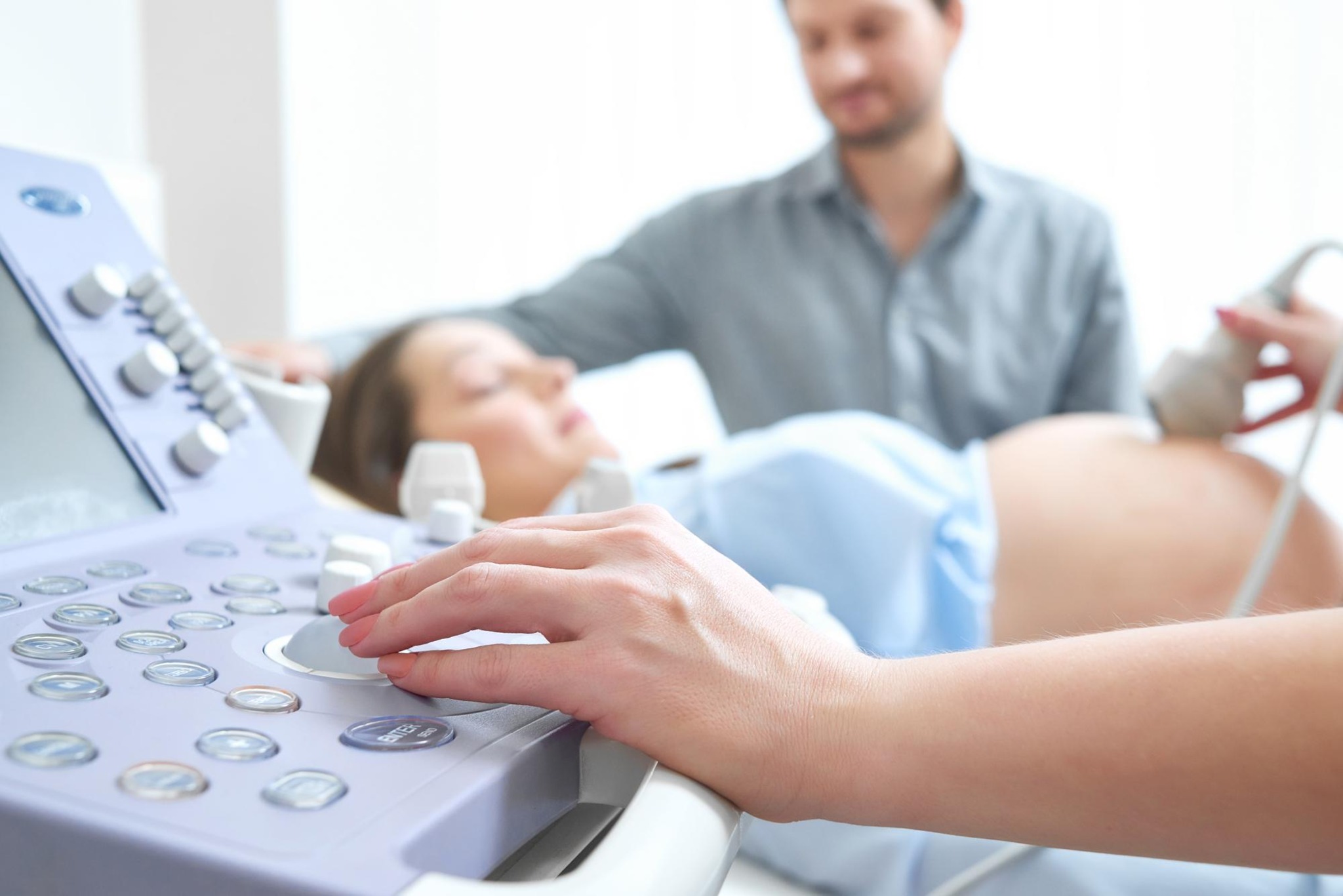
What is a morphological ultrasound?
A morphological ultrasound, or morphological ultrasonography, is an imaging exam performed during pregnancy to evaluate the baby's development in detail. Through sound waves, we can create images of the fetus's structures and organs.
This exam is more detailed than a regular ultrasound and its main purpose is to identify possible malformations and growth abnormalities in the baby. Furthermore, it is essential for determining if the pregnancy presents any risk factors, allowing medical monitoring to be adjusted to provide the best care for both mother and baby.
Two important moments: the first and second trimester morphological ultrasounds.
Many people don't know that there are two types of morphological ultrasound, each with specific purposes and performed at different times during pregnancy. I'll explain the differences:
First Trimester Morphological Ultrasound
This examination is performed between the 12th and 14th week of gestation. It allows for an initial assessment of the baby's anatomy and helps identify genetic syndromes, such as Down syndrome, as well as congenital malformations.
Another important point is the risk assessment for conditions such as pre-eclampsia, which is high blood pressure during pregnancy. This information is valuable because it allows for early interventions to be carried out if necessary.
Second Trimester Morphological Ultrasound
The second-trimester ultrasound is performed between the 20th and 24th week of gestation. At this stage, the baby is more developed, allowing for an even more detailed analysis of its organs and structures, such as the heart, brain, kidneys, and limbs.
This ultrasound is especially useful for detecting serious malformations and assisting in birth planning or potential medical interventions.
What can we identify with a morphological ultrasound?
This exam offers a complete overview of the baby's health and development. Among the information we can obtain are:
- Fetal malformations: Identifying developmental anomalies in babies, such as problems with the heart, brain, kidneys, and even limbs.
- Fetal monitoring: Monitoring fetal growth, verifying if development is as expected for gestational age.
- Baby's position: During the examination, it is possible to check the baby's position in the uterus, which helps in planning the type of delivery.
- Amount of amniotic fluid: Checking the amount of amniotic fluid is essential for the baby's well-being.
The impact of missing a pregnancy test
If a morphological ultrasound is not performed, many important conditions may go unnoticed. For example, malformations or alterations in the baby's growth may not be identified, making it difficult to properly plan the delivery and prenatal care.
Furthermore, without this examination, risk factors during pregnancy may not be detected early, which can negatively impact the health of both the baby and the mother.
Are there risks involved in performing a morphological ultrasound?
No, this exam is completely safe. The morphological ultrasound is a non-invasive procedure, meaning it doesn't involve any cutting or insertion of instruments into the body. It uses only sound waves, without radiation, making it safe for both mother and baby.
Myths about morphological ultrasound
1) Morphological ultrasound can cause harm to the baby.
Myth: Morphological ultrasound is completely safe, does not harm the baby, and involves no radiation.
2) The examination is painful.
Myth: The procedure is painless. In fact, it's a quick and simple examination in which a gel is applied to the abdomen to facilitate the transmission of sound waves.
3) The test can detect all diseases.
Myth: Although morphological ultrasound is very effective, it cannot detect all diseases or complications. Other tests may be necessary, as directed by a doctor.
As I explained, this test is safe, effective, and should be performed at the appropriate time so that we can monitor each stage of your pregnancy in a calm and safe manner.
If you have any questions or need guidance, I'm here to help.